
The 9th Edition of Radiation Protection in Medical Radiography provides a comprehensive guide to understanding radiation safety principles and practices in medical imaging․ It covers the safe use of ionizing radiation, its biological effects, and modern techniques to minimize exposure, ensuring patient and staff safety while maintaining image quality․
Overview of the 9th Edition
The 9th Edition of Radiation Protection in Medical Radiography offers a detailed exploration of radiation safety, blending foundational and advanced concepts․ It features full-color illustrations, updated content on radiation physics, and practical applications in modern imaging modalities․ The edition emphasizes minimizing radiation exposure while maintaining diagnostic image quality, making it a valuable resource for radiography students and professionals seeking to enhance their understanding of radiation protection principles and practices․
Importance of Radiation Safety in Medical Imaging
Radiation safety is fundamental to protecting patients, staff, and the public from the risks of ionizing radiation․ It ensures the safe use of imaging technologies, balancing diagnostic benefits with exposure risks․ Proper safety protocols minimize radiation-induced biological effects, reducing long-term health risks․ This approach promotes ethical imaging practices, enhancing patient trust and overall well-being while maintaining image quality for accurate diagnoses․
Basic Principles of Radiation Protection
Radiation protection relies on minimizing exposure through time, distance, and shielding, ensuring safety for patients and staff while using ionizing radiation in medical imaging․
Key Concepts: ALARA, Time, Distance, and Shielding
ALARA (As Low As Reasonably Achievable) is a cornerstone of radiation protection, emphasizing minimizing exposure․ Time reduction lowers dose, while distance leverages the inverse square law to decrease exposure․ Shielding with lead or other materials blocks radiation, protecting both patients and staff․ These principles ensure safe use of ionizing radiation in medical imaging, balancing image quality with safety․ They are fundamental in all radiographic practices․
Radiation Physics and Its Role in Medical Radiography
Radiation physics is fundamental to medical radiography, governing the interaction of X-rays with tissue․ Understanding beam composition, attenuation, and scattering enables precise image formation․ Physics principles guide equipment design and exposure settings, ensuring optimal image quality while minimizing dose․ This knowledge is essential for selecting appropriate techniques and energies, balancing diagnostic needs with patient safety in radiographic imaging․
Biological Effects of Ionizing Radiation
Ionizing radiation affects cells and systems, causing damage at the molecular and cellular levels․ Understanding these effects is crucial for minimizing risks and ensuring safe practices in medical imaging․
Cellular and Systemic Effects of Radiation Exposure
Radiation exposure can cause damage to cellular DNA, leading to mutations and potentially harmful effects․ At the cellular level, ionizing radiation disrupts molecular structures, affecting normal function․ Systemically, prolonged or high doses of radiation can result in organ damage, increased cancer risk, and immune system suppression․ Understanding these effects is critical for developing strategies to minimize exposure and protect both patients and professionals in medical imaging settings․
Radiobiology and Risk Assessment
Radiobiology examines the effects of ionizing radiation on living cells and tissues, focusing on DNA damage and repair mechanisms․ Risk assessment quantifies these effects, determining the likelihood of harm from radiation exposure․ Understanding dose-response relationships helps establish safe limits for radiation use in medical imaging․ This knowledge is vital for balancing diagnostic benefits with potential risks, ensuring optimal patient care and safety in radiographic procedures․
Safety Measures in Medical Radiography
Safety measures in medical radiography include implementing protocols to minimize radiation exposure, using personal protective equipment, and adhering to regulatory guidelines to ensure patient and staff protection․
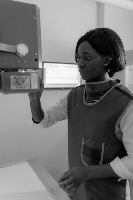
Personal Protective Equipment (PPE) for Radiographers
Personal protective equipment (PPE) is essential for radiographers to minimize radiation exposure․ This includes lead aprons, thyroid collars, gloves, and eyewear․ Proper fit and regular inspection ensure effectiveness․ PPE is designed to shield against scattered radiation, protecting sensitive areas․ Its use is mandated by safety protocols to safeguard health and comply with regulatory standards, ensuring both patient and staff safety during imaging procedures․
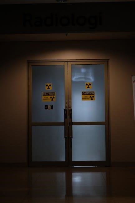
Design and Shielding of Radiographic Facilities
Effective design and shielding of radiographic facilities are critical for radiation protection; Primary and secondary barriers, such as lead-lined walls and ceilings, minimize radiation exposure․ Materials like lead and gypsum are used to absorb or block scattered radiation․ Facility design ensures optimal workflow while maintaining safety․ Shielding requirements are tailored to the type and energy of radiation equipment, adhering to NCRP and ICRP guidelines to protect patients, staff, and the public from unnecessary radiation exposure․
Regulatory and Advisory Limits for Radiation Exposure
NCRP and ICRP Guidelines for Radiation Protection
The National Council on Radiation Protection (NCRP) and International Commission on Radiological Protection (ICRP) establish exposure limits and safety standards․ These guidelines ensure radiation doses remain as low as reasonably achievable (ALARA), balancing diagnostic benefits with risks․ Annual dose limits for workers and the public are specified, emphasizing shielding, time, and distance principles to minimize exposure in medical radiography, aligning with global best practices for radiation protection․
The National Council on Radiation Protection (NCRP) and the International Commission on Radiological Protection (ICRP) provide comprehensive guidelines to ensure safe radiation practices․ These organizations establish dose limits for workers and the public, emphasizing the ALARA principle (As Low As Reasonably Achievable)․ Annual exposure limits are set to minimize risks, with specific thresholds for radiographers and patients․ The guidelines also recommend shielding, distance, and time management to reduce exposure, aligning with modern radiography practices to optimize safety and diagnostic quality effectively․
Implementation of Safety Standards in Clinical Practice
Implementing safety standards in clinical practice is crucial for minimizing radiation exposure․ The 9th Edition emphasizes adhering to ALARA principles, proper use of PPE, and regular equipment maintenance․ These practices reduce radiation exposure risks, ensuring both patient and staff safety․ Continuous education and quality control measures further enhance compliance, optimizing image quality while maintaining safety protocols effectively․

Patient Protection in Medical Radiography
The 9th Edition focuses on minimizing radiation exposure to patients through optimized imaging techniques and adherence to the ALARA principle, ensuring safety while maintaining diagnostic image quality․
Minimizing Patient Dose During Imaging Procedures
Minimizing radiation exposure to patients is critical in medical radiography․ The 9th Edition emphasizes techniques such as adjusting exposure factors, using digital imaging, and applying the ALARA principle․ Proper patient positioning, shielding, and limiting the field of view reduce unnecessary radiation․ Modern technologies, like dose-reduction software, further optimize patient safety․ These strategies ensure diagnostic image quality while minimizing the risks associated with ionizing radiation, aligning with best practices in radiation protection and patient care․
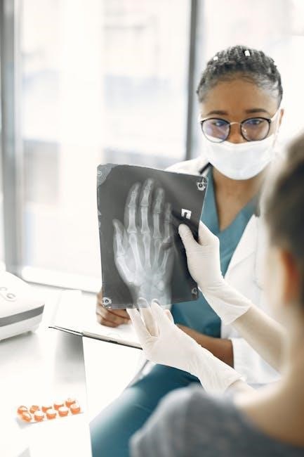
Communication and Education for Patient Safety
Effective communication and education are vital for ensuring patient safety in radiography․ Informing patients about procedures and risks fosters trust and cooperation․ Radiographers should explain the importance of remaining still and following instructions to minimize radiation exposure․ Education also involves discussing the benefits and risks of imaging, ensuring patients understand the necessity of the procedure․ Clear communication helps reduce anxiety, enhances safety, and supports optimal imaging outcomes while minimizing radiation risks․

Equipment and Technology in Radiation Protection
Modern imaging technologies, such as digital radiography and computed radiography, play a crucial role in minimizing radiation exposure while maintaining high-quality images for accurate diagnostics․
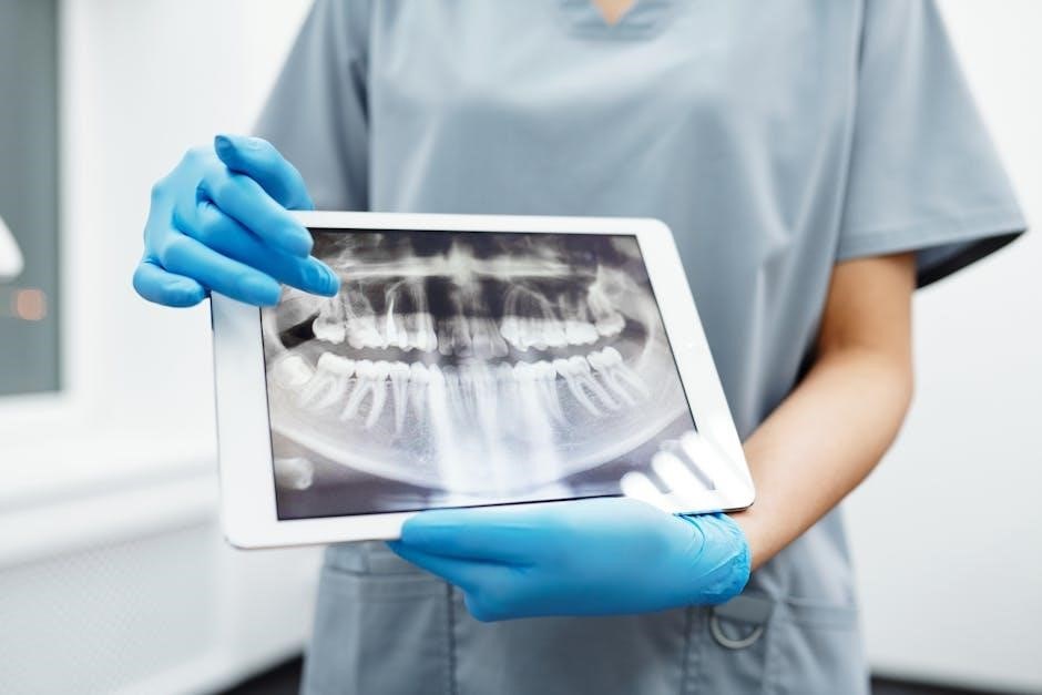
Modern Imaging Modalities and Their Safety Features
Modern imaging modalities, such as digital radiography and computed radiography, incorporate advanced safety features to minimize radiation exposure․ These technologies enable precise dose control, reducing patient exposure while maintaining image quality․ Digital systems also offer tools for dose monitoring and optimization, ensuring compliance with radiation safety standards․ Such advancements highlight the integration of technology in balancing diagnostic accuracy with patient protection, aligning with the principles outlined in the 9th Edition of Radiation Protection in Medical Radiography․
Quality Control and Maintenance of Radiographic Equipment
Regular quality control and maintenance of radiographic equipment are crucial for ensuring optimal performance and radiation safety․ The 9th Edition emphasizes routine testing, calibration, and servicing to prevent equipment malfunctions․ Proper maintenance minimizes radiation exposure risks, ensuring accurate image production while adhering to safety standards․ This proactive approach guarantees reliable operation, protecting both patients and staff from unnecessary radiation risks in medical imaging environments․

Future Directions in Radiation Protection
Future directions in radiation protection involve leveraging emerging technologies such as AI and advanced shielding to enhance safety and efficiency in medical radiography, ensuring optimal patient care․
Emerging Technologies and Their Impact on Safety
Emerging technologies like artificial intelligence (AI) and advanced imaging modalities are revolutionizing radiation protection․ AI optimizes dose reduction while maintaining image quality, minimizing patient exposure․ Innovations in shielding materials and real-time radiation monitoring systems enhance safety for both patients and radiographers․ Additionally, 3D printing enables customized shielding solutions, further reducing exposure risks․ These advancements underscore the potential for safer, more efficient practices in medical radiography, aligning with modern safety standards and patient care goals;
Continuous Education and Training for Radiographers
Continuous education and training are essential for radiographers to stay updated on radiation protection best practices․ The 9th Edition emphasizes the importance of lifelong learning, offering structured programs and resources to enhance skills and knowledge․ Regular training ensures adherence to safety standards, fosters professional development, and promotes optimal patient care․ By prioritizing education, radiographers can effectively integrate new technologies and safety protocols, maintaining high standards in radiation protection and medical imaging․